In cardiac physiology, cardiac output (CO), also known as heart output and often denoted by the symbols , , or ,[2] is the volumetric flow rate of the heart's pumping output: that is, the volume of blood being pumped by a single ventricle of the heart, per unit time (usually measured per minute). Cardiac output (CO) is the product of the heart rate (HR), i.e. the number of heartbeats per minute (bpm), and the stroke volume (SV), which is the volume of blood pumped from the left ventricle per beat; thus giving the formula:
Values for cardiac output are usually denoted as L/min. For a healthy individual weighing 70 kg, the cardiac output at rest averages about 5 L/min; assuming a heart rate of 70 beats/min, the stroke volume would be approximately 70 mL.
Because cardiac output is related to the quantity of blood delivered to various parts of the body, it is an important component of how efficiently the heart can meet the body's demands for the maintenance of adequate tissue perfusion. Body tissues require continuous oxygen delivery which requires the sustained transport of oxygen to the tissues by systemic circulation of oxygenated blood at an adequate pressure from the left ventricle of the heart via the aorta and arteries. Oxygen delivery (DO2 mL/min) is the resultant of blood flow (cardiac output CO) times the blood oxygen content (CaO2). Mathematically this is calculated as follows: oxygen delivery = cardiac output × arterial oxygen content, giving the formula:
With a resting cardiac output of 5 L/min, a 'normal' oxygen delivery is around 1 L/min. The amount/percentage of the circulated oxygen consumed (VO2) per minute through metabolism varies depending on the activity level but at rest is circa 25% of the DO2. Physical exercise requires a higher than resting-level of oxygen consumption to support increased muscle activity. In the case of heart failure, actual CO may be insufficient to support even simple activities of daily living; nor can it increase sufficiently to meet the higher metabolic demands stemming from even moderate exercise.
Cardiac output is a global blood flow parameter of interest in hemodynamics, the study of the flow of blood. The factors affecting stroke volume and heart rate also affect cardiac output. The figure at the right margin illustrates this dependency and lists some of these factors. A detailed hierarchical illustration is provided in a subsequent figure.
There are many methods of measuring CO, both invasively and non-invasively; each has advantages and drawbacks as described below.

The function of the heart is to drive blood through the circulatory system in a cycle that delivers oxygen, nutrients and chemicals to the body's cells and removes cellular waste. Because it pumps out whatever blood comes back into it from the venous system, the quantity of blood returning to the heart effectively determines the quantity of blood the heart pumps out – its cardiac output, Q. Cardiac output is classically defined alongside stroke volume (SV) and the heart rate (HR) as:[citation needed]
In standardizing what CO values are considered to be within normal range independent of the size of the subject's body, the accepted convention is to further index equation (1) using body surface area (BSA), giving rise to the Cardiac index (CI). This is detailed in equation (2) below.
There are a number of clinical methods to measure cardiac output, ranging from direct intracardiac catheterization to non-invasive measurement of the arterial pulse. Each method has advantages and drawbacks. Relative comparison is limited by the absence of a widely accepted "gold standard" measurement. Cardiac output can also be affected significantly by the phase of respiration – intra-thoracic pressure changes influence diastolic filling and therefore cardiac output. This is especially important during mechanical ventilation, in which cardiac output can vary by up to 50% across a single respiratory cycle.[citation needed] Cardiac output should therefore be measured at evenly spaced points over a single cycle or averaged over several cycles.[citation needed]
Invasive methods are well accepted, but there is increasing evidence that these methods are neither accurate nor effective in guiding therapy. Consequently, the focus on development of non-invasive methods is growing.[5][6][7]

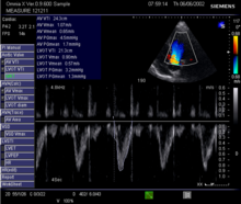
This method uses ultrasound and the Doppler effect to measure cardiac output. The blood velocity through the heart causes a Doppler shift in the frequency of the returning ultrasound waves. This shift can then be used to calculate flow velocity and volume, and effectively cardiac output, using the following equations:[citation needed]
where:
Being non-invasive, accurate and inexpensive, Doppler ultrasound is a routine part of clinical ultrasound; it has high levels of reliability and reproducibility, and has been in clinical use since the 1960s.[citation needed]
Echocardiography is a non-invasive method of quantifying cardiac output using ultrasound. Two-dimensional (2D) ultrasound and Doppler measurements are used together to calculate cardiac output. 2D measurement of the diameter (d) of the aortic annulus allows calculation of the flow cross-sectional area (CSA), which is then multiplied by the VTI of the Doppler flow profile across the aortic valve to determine the flow volume per beat (stroke volume, SV). The result is then multiplied by the heart rate (HR) to obtain cardiac output. Although used in clinical medicine, it has a wide test-retest variability.[8] It is said to require extensive training and skill, but the exact steps needed to achieve clinically adequate precision have never been disclosed. 2D measurement of the aortic valve diameter is one source of noise; others are beat-to-beat variation in stroke volume and subtle differences in probe position. An alternative that is not necessarily more reproducible is the measurement of the pulmonary valve to calculate right-sided CO. Although it is in wide general use, the technique is time-consuming and is limited by the reproducibility of its component elements. In the manner used in clinical practice, precision of SV and CO is of the order of ±20%.[citation needed]
Ultrasonic Cardiac Output Monitor (USCOM) uses continuous wave Doppler to measure the Doppler flow profile VTI. It uses anthropometry to calculate aortic and pulmonary valve diameters and CSAs, allowing right-sided and left-sided Q measurements. In comparison to the echocardiographic method, USCOM significantly improves reproducibility and increases sensitivity of the detection of changes in flow. Real-time, automatic tracing of the Doppler flow profile allows beat-to-beat right-sided and left-sided Q measurements, simplifying operation and reducing the time of acquisition compared to conventional echocardiography. USCOM has been validated from 0.12 L/min to 18.7 L/min[9] in new-born babies,[10] children[11] and adults.[12] The method can be applied with equal accuracy to patients of all ages for the development of physiologically rational haemodynamic protocols. USCOM is the only method of cardiac output measurement to have achieved equivalent accuracy to the implantable flow probe.[13] This accuracy has ensured high levels of clinical use in conditions including sepsis, heart failure and hypertension.[14][15][16]

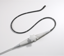
The Transoesophageal Doppler includes two main technologies; transoesophageal echocardiogram—which is primarily used for diagnostic purposes, and oesophageal Doppler monitoring—which is primarily used for the clinical monitoring of cardiac output. The latter uses continuous wave Doppler to measure blood velocity in the descending thoracic aorta. An ultrasound probe is inserted either orally or nasally into the oesophagus to mid-thoracic level, at which point the oesophagus lies alongside the descending thoracic aorta. Because the transducer is close to the blood flow, the signal is clear. The probe may require re-focussing to ensure an optimal signal. This method has good validation, is widely used for fluid management during surgery with evidence for improved patient outcome,[17][18][19][20][21][22][23][24] and has been recommended by the UK's National Institute for Health and Clinical Excellence (NICE).[25] Oesophageal Doppler monitoring measures the velocity of blood and not true Q, therefore relies on a nomogram[26] based on patient age, height and weight to convert the measured velocity into stroke volume and cardiac output. This method generally requires patient sedation and is accepted for use in both adults and children.[citation needed]
Pulse pressure (PP) methods measure the pressure in an artery over time to derive a waveform and use this information to calculate cardiac performance. However, any measure from the artery includes changes in pressure associated with changes in arterial function, for example compliance and impedance. Physiological or therapeutic changes in vessel diameter are assumed to reflect changes in Q. PP methods measure the combined performance of the heart and the blood vessels, thus limiting their application for measurement of Q. This can be partially compensated for by intermittent calibration of the waveform to another Q measurement method then monitoring the PP waveform. Ideally, the PP waveform should be calibrated on a beat-to-beat basis. There are invasive and non-invasive methods of measuring PP.[citation needed]
In 1967, the Czech physiologist Jan Peňáz invented and patented the volume clamp method of measuring continuous blood pressure. The principle of the volume clamp method is to dynamically provide equal pressures, on either side of an artery wall. By clamping the artery to a certain volume, inside pressure—intra-arterial pressure—balances outside pressure—finger cuff pressure. Peñáz decided the finger was the optimal site to apply this volume clamp method. The use of finger cuffs excludes the device from application in patients without vasoconstriction, such as in sepsis or in patients on vasopressors.[citation needed]
In 1978, scientists at BMI-TNO, the research unit of Netherlands Organisation for Applied Scientific Research at the University of Amsterdam, invented and patented a series of additional key elements that make the volume clamp work in clinical practice. These methods include the use of modulated infrared light in the optical system inside the sensor, the lightweight, easy-to-wrap finger cuff with velcro fixation, a new pneumatic proportional control valve principle, and a set point strategy for the determining and tracking the correct volume at which to clamp the finger arteries—the Physiocal system. An acronym for physiological calibration of the finger arteries, this Physiocal tracker was found to be accurate, robust and reliable.[citation needed]
The Finapres methodology was developed to use this information to calculate arterial pressure from finger cuff pressure data. A generalised algorithm to correct for the pressure level difference between the finger and brachial sites in patients was developed. This correction worked under all of the circumstances it was tested in—even when it was not designed for it—because it applied general physiological principles. This innovative brachial pressure waveform reconstruction method was first implemented in the Finometer, the successor of Finapres that BMI-TNO introduced to the market in 2000.[citation needed]
The availability of a continuous, high-fidelity, calibrated blood pressure waveform opened up the perspective of beat-to-beat computation of integrated haemodynamics, based on two notions: pressure and flow are inter-related at each site in the arterial system by their so-called characteristic impedance. At the proximal aortic site, the 3-element Windkessel model of this impedance can be modelled with sufficient accuracy in an individual patient with known age, gender, height and weight. According to comparisons of non-invasive peripheral vascular monitors, modest clinical utility is restricted to patients with normal and invariant circulation.[27]
Invasive PP monitoring involves inserting a manometer pressure sensor into an artery—usually the radial or femoral artery—and continuously measuring the PP waveform. This is generally done by connecting the catheter to a signal processing device with a display. The PP waveform can then be analysed to provide measurements of cardiovascular performance. Changes in vascular function, the position of the catheter tip or damping of the pressure waveform signal will affect the accuracy of the readings. Invasive PP measurements can be calibrated or uncalibrated.[citation needed]
PiCCO (PULSION Medical Systems AG, Munich, Germany) and PulseCO (LiDCO Ltd, London, England) generate continuous Q by analysing the arterial PP waveform. In both cases, an independent technique is required to provide calibration of continuous Q analysis because arterial PP analysis cannot account for unmeasured variables such as the changing compliance of the vascular bed. Recalibration is recommended after changes in patient position, therapy or condition.[citation needed]
In PiCCO, transpulmonary thermodilution, which uses the Stewart-Hamilton principle but measures temperatures changes from central venous line to a central arterial line, i.e., the femoral or axillary arterial line, is used as the calibrating technique. The Q value derived from cold-saline thermodilution is used to calibrate the arterial PP contour, which can then provide continuous Q monitoring. The PiCCO algorithm is dependent on blood pressure waveform morphology (mathematical analysis of the PP waveform), and it calculates continuous Q as described by Wesseling and colleagues.[28] Transpulmonary thermodilution spans right heart, pulmonary circulation and left heart, allowing further mathematical analysis of the thermodilution curve and giving measurements of cardiac filling volumes (GEDV), intrathoracic blood volume and extravascular lung water. Transpulmonary thermodilution allows for less invasive Q calibration but is less accurate than PA thermodilution and requires a central venous and arterial line with the accompanied infection risks.[citation needed]
In LiDCO, the independent calibration technique is lithium chloride dilution using the Stewart-Hamilton principle. Lithium chloride dilution uses a peripheral vein and a peripheral arterial line. Like PiCCO, frequent calibration is recommended when there is a change in Q.[29] Calibration events are limited in frequency because they involve the injection of lithium chloride and can be subject to errors in the presence of certain muscle relaxants. The PulseCO algorithm used by LiDCO is based on pulse power derivation and is not dependent on waveform morphology.[citation needed]
FloTrac/Vigileo (Edwards Lifesciences) is an uncalibrated, haemodynamic monitor based on pulse contour analysis. It estimates cardiac output (Q) using a standard arterial catheter with a manometer located in the femoral or radial artery. The device consists of a high-fidelity pressure transducer, which, when used with a supporting monitor (Vigileo or EV1000 monitor), derives left-sided cardiac output (Q) from a sample of arterial pulsations. The device uses an algorithm based on the Frank–Starling law of the heart, which states pulse pressure (PP) is proportional to stroke volume (SV). The algorithm calculates the product of the standard deviation of the arterial pressure (AP) wave over a sampled period of 20 seconds and a vascular tone factor (Khi, or χ) to generate stroke volume. The equation in simplified form is: , or, . Khi is designed to reflect arterial resistance; compliance is a multivariate polynomial equation that continuously quantifies arterial compliance and vascular resistance. Khi does this by analyzing the morphological changes of arterial pressure waveforms on a bit-by-bit basis, based on the principle that changes in compliance or resistance affect the shape of the arterial pressure waveform. By analyzing the shape of said waveforms, the effect of vascular tone is assessed, allowing the calculation of SV. Q is then derived using equation (1). Only perfused beats that generate an arterial waveform are counted for in HR.[citation needed]
This system estimates Q using an existing arterial catheter with variable accuracy. These arterial monitors do not require intracardiac catheterisation from a pulmonary artery catheter. They require an arterial line and are therefore invasive. As with other arterial waveform systems, the short set-up and data acquisition times are benefits of this technology. Disadvantages include its inability to provide data regarding right-sided heart pressures or mixed venous oxygen sa